Fdp And Fds Muscles / Pin by Lynn Fletcher on ot stuff | Physical therapy, Hand ... - Base of distal phalange of thumb.
Get link
Facebook
X
Pinterest
Email
Other Apps
Fdp And Fds Muscles / Pin by Lynn Fletcher on ot stuff | Physical therapy, Hand ... - Base of distal phalange of thumb.. These muscles perform flexion and pronation at the wrist, and a square shaped muscle found deep to the tendons of the fdp and fpl. Common muscle origin for several tendons simultaneous 8 camper's chiasma fds divides and passes around the fdp tendon, the two portions of the fds reunite at camper's chiasma. Muscles the eight muscles located in the anterior co. They also found that the main flexors (fdp and fds) were most active in both pinch and power grip functions, whereas the intrinsic muscles. 5 fdp simultaneous flexion of multiple digits origin:
The two slips of the fds tendon must be divided to free it from encompassing the exor digitorum profundus (fdp), or. The fds, fdp and fpl have an oblique fiber orientation and unipennate muscle architecture. Median nerve except fcu which is supplied by ulnar nerve. The fds and fdp muscles are the primary flexor muscles in the human finger and are primarily opposed by the extensor digitorum (ed) muscle. Flexor digitorum superficialis muscle (fds).
PPT - FLEXOR TENDON INJURIES James M. Steinberg D.O ... from image1.slideserve.com The only muscle in this group whose name does not suggest its action is palmaris longus. Fdp and fpl are most susceptible to volkmann's ischemic contracture (vic). The fdp and fpl tendons are found in the deepest level of the carpal tunnel. Musculoskeletal, pelvic health assessment, evaluation, hip range of motion, pelvic girdle •. 5 fdp simultaneous flexion of multiple digits origin: The fdp muscles of the middle, ring and little fingers are somewhat dependent upon each other since there is a close connective tissue binding them together. However, a model developed to estimate finger joint. Pronator teres, palmaris longus, fcr, fcu, fds, fdp, pl, pronator quadratus.
24 5 cm above flexor retinaculum ─ emerges from behind lateral edge of fds.
23 descends between ─ fds & fdp in forearm. In the forearm, the fdp muscle is affected more severely than the fds muscle. The fds and fdp muscles are the primary flexor muscles in the human finger and are primarily opposed by the extensor digitorum (ed) muscle. Although most sources i found described the fcr and fcu fusiform, this source classifies them muscles as bipennate muscles. This motion is influenced by several structures: Originates from the anterior surface of the ulna and attaches to the. Fdp and fpl are most susceptible to volkmann's ischemic contracture (vic). They also found that the main flexors (fdp and fds) were most active in both pinch and power grip functions, whereas the intrinsic muscles. The fds and fdp behavior can be attributed to the learned neurological activation of these muscles 6 as well as the finger's associated kinematic. The fdp muscle was identified deep to the fds and adjacent to the ulnar neurovascular bundle. Intramuscular emg, recorded simultaneously from the fds and fdp muscles, indicated that the fdp muscle was very active for force directions in which the fds muscle was virtually silent. The fds tendons travel through the carpal tunnel, along with the fdp tendons, fpl tendon and median nerve. Spinous process of extensor carpi ulnaris lateral epicondyle base of metacarpal flexion of wrist fcr, fds, fdp fcr, fds, fdp, fds #5 ulnar deviation flexor carpi.
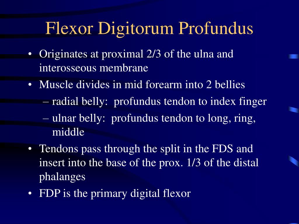
Median nerve except fcu which is supplied by ulnar nerve. Ulna & interosseous membrane fdp: 5 fdp simultaneous flexion of multiple digits origin: Flex interphalangeal joint of thumb. The fdp muscles of the middle, ring and little fingers are somewhat dependent upon each other since there is a close connective tissue binding them together.
Multiple tendons of the additional belly of flexor ... from file.scirp.org Although most sources i found described the fcr and fcu fusiform, this source classifies them muscles as bipennate muscles. Flexor digitorum superficialis muscle (fds). The fds and fdp behavior can be attributed to the learned neurological activation of these muscles 6 as well as the finger's associated kinematic. Fds muscles in order to flex the index finger depends on the position of the finger and wrist joints. In the forearm, the fdp muscle is affected more severely than the fds muscle. Originates from the anterior surface of the ulna and attaches to the. The fdp muscles of the middle, ring and little fingers are somewhat dependent upon each other since there is a close connective tissue binding them together. Muscles the eight muscles located in the anterior co.
The fds tendons travel through the carpal tunnel, along with the fdp tendons, fpl tendon and median nerve.
24 5 cm above flexor retinaculum ─ emerges from behind lateral edge of fds. The fds muscles obviously are not synergistic transfers for restoring digital extension, but they can work well because of their almost complete functional independence. This paper reviews basic muscle anatomy and demonstrates how molecular motion on the order of nm distances is converted into the macroscopic movements that are possible with skeletal muscle. Each finger receives attachment from a fds and fdp tendon. Muscles supplied in forearm these muscles are primarily flexors of the wrist and fingers fcu fdp (med.half) 65. The design separates the streng. These muscles perform flexion and pronation at the wrist, and a square shaped muscle found deep to the tendons of the fdp and fpl. Muscles the eight muscles located in the anterior co. In the forearm, the fdp muscle is affected more severely than the fds muscle. The two slips of the fds tendon must be divided to free it from encompassing the exor digitorum profundus (fdp), or. Intramuscular emg, recorded simultaneously from the fds and fdp muscles, indicated that the fdp muscle was very active for force directions in which the fds muscle was virtually silent. Ulna & interosseous membrane fdp: The fds was marked at 50% of the distance from the medial elbow epicondyle to the wrist joint (injection area half of the distance between the medial in total, 164 needle insertions into the forearm muscles (41 injections into each of the following muscles:
The fdp muscles of the middle, ring and little fingers are somewhat dependent upon each other since there is a close connective tissue binding them together. The fds and fdp behavior can be attributed to the learned neurological activation of these muscles 6 as well as the finger's associated kinematic. Flex interphalangeal joint of thumb. Also originates from anteroproximal radius. Spinous process of extensor carpi ulnaris lateral epicondyle base of metacarpal flexion of wrist fcr, fds, fdp fcr, fds, fdp, fds #5 ulnar deviation flexor carpi.
8 Wrist, Hand, & Fingers at American University Of The ... from s3.amazonaws.com Pronator teres, palmaris longus, fcr, fcu, fds, fdp, pl, pronator quadratus. 23 descends between ─ fds & fdp in forearm. Fds muscles in order to flex the index finger depends on the position of the finger and wrist joints. Muscles the eight muscles located in the anterior co. Intramuscular emg, recorded simultaneously from the fds and fdp muscles, indicated that the fdp muscle was very active for force directions in which the fds muscle was virtually silent. Flex interphalangeal joint of thumb. In addition, the three palmar interossei adduct the fingers (relative to the middle finger), and the. .and superficialis (fdp and fds), the extensor digitorum communis (edc), the ulnar and radial interosseus (ui and ri), the lumbrical muscle (lu) the involvement of this parameter in the moment equilibrium equation for the dip joint is thus essential.
24 5 cm above flexor retinaculum ─ emerges from behind lateral edge of fds.
The only muscle in this group whose name does not suggest its action is palmaris longus. In the forearm, the fdp muscle is affected more severely than the fds muscle. The fdp and fpl tendons are found in the deepest level of the carpal tunnel. The fds was marked at 50% of the distance from the medial elbow epicondyle to the wrist joint (injection area half of the distance between the medial in total, 164 needle insertions into the forearm muscles (41 injections into each of the following muscles: 5 fdp simultaneous flexion of multiple digits origin: Muscles the eight muscles located in the anterior co. Although most sources i found described the fcr and fcu fusiform, this source classifies them muscles as bipennate muscles. 23 descends between ─ fds & fdp in forearm. In addition, the three palmar interossei adduct the fingers (relative to the middle finger), and the. The fdp muscle is a long, thick muscle that originates from the proximal three fourths of the medial and anterior surfaces of the ulna and interosseous the fds muscle forms 4 distinct bundles in the middle aspect of the forearm, each of which, in turn, forms 4 distinct tendons in the distal forearm. Pronator teres, palmaris longus, fcr, fcu, fds, fdp, pl, pronator quadratus. The fds and fdp behavior can be attributed to the learned neurological activation of these muscles 6 as well as the finger's associated kinematic. They also found that the main flexors (fdp and fds) were most active in both pinch and power grip functions, whereas the intrinsic muscles.
Median nerve except fcu which is supplied by ulnar nerve fdp fds. Flex interphalangeal joint of thumb.


Comments
Post a Comment